
Laboratory Equipment & Services Information System
by Centralized Laboratory Management Office (CeLMO)
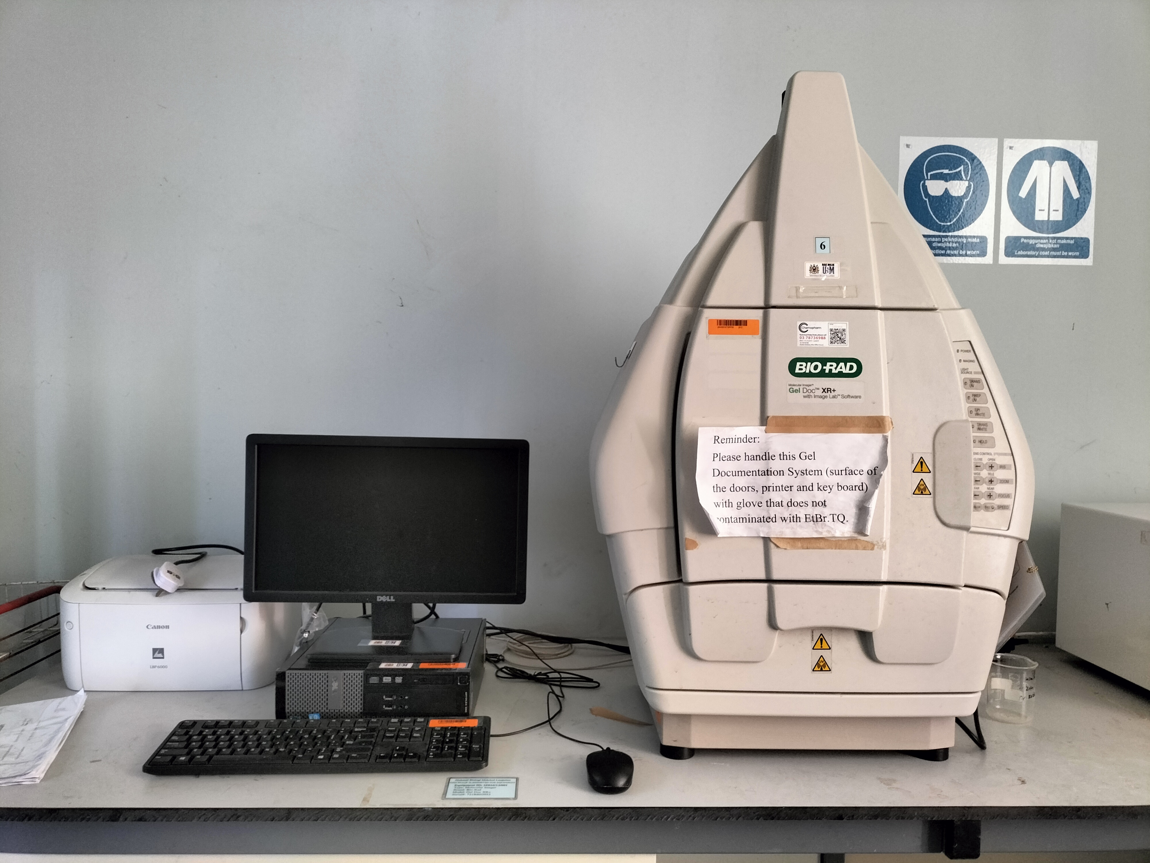
Gel Doc™ XR+ and ChemiDoc™ XRS+ gel documentation systems offer high performance and ease of use. They both contain a charge coupled device (CCD) camera to capture images in real time and allow you to accurately position your sample and generate optimized image data. The Gel Doc XR+ and ChemiDoc XRS+ systems use the same light tight enclosure (the universal hood), which contains built-in UV and white light illumination, but different CCD cameras. Both systems feature dynamic flat fielding technology for superior image uniformity and accurate quantitation. Bio-Rad® Image Lab™ software controls image capture and optimization for your selected applications, analyzes results, and produces reports based on your specified output, all in a single workflow.
The system accommodates a wide array of samples, from large hand-cast polyacrylamide gels to small Ready Agarose Gels and various blots. The system is an ideal accompaniment to PCR, purification, and electrophoresis systems, enabling image analysis and documentation of restriction digests, amplified nucleic acids, genetic fingerprinting, RFLPs, and protein purification and characterization Gel Doc XR+ Imager Workflow Following are the basic steps to acquiring, analyzing, and archiving an image using the Gel Doc XR+ system and Image Lab software: 1. Select an existing protocol or customize a new one. 2. Position the sample to be imaged. 3. Run a selected protocol. 4. View the displayed results. 5. Optimize the analysis. 6. Generate a report. 7. Save or export the results
The Gel Doc XR+ System is based on CCD high-resolution, high-sensitivity detection technology and modular options to accommodate a wide range of samples and support multiple detection methods including fluorescence and colorimetric detection. The Gel Doc XR+ System is controlled by Image Lab Software to optimize imager performance for fast, integrated, and automated image capture and analysis of various samples.
- Manufacturer
- Brand
- BIO RAD
- Model
- GEL DOC XR+
- Year Manufactured
- Year Procured
- 2012
- Department
- PUSAT PENGAJIAN SAINS KESIHATAN
- Location
- Ppsk-blok C > Satu> Makmal Biologi Molekul>bilik 4
- Date Registered LESIS
- 04/03/2024
- Category
- Research Equipment
- Function
- Booking,
- Category
- Staff operated
- Equipment Status
- Good
Person In-Charge
