

Laboratory Equipment & Services Information System
by Centralized Laboratory Management Office (CeLMO)
> High vacuum mode (chamber vacuum pressure < 6 e-4 Pa; nominal 1.0 nm electron beam resolution at 30 kV) used for high-resolution imaging and microanalyses of conductive samples in both secondary electron (SE) and backscattered (BSE) modes. > Low vacuum mode (chamber vacuum pressure 10 – 130 Pa; nominal 1.4 nm electron beam resolution at 30 kV) for high-resolution imaging and microanalyses of non-conductive samples in both secondary electron and backscatter modes. > Extended low vacuum mode ESEM (chamber vacuum pressure 10 – 4000 Pa; nominal 1.4 nm electron beam resolution at 30 kV) for charge-free imaging and microanalysis of non-conductive and/or hydrated specimens.
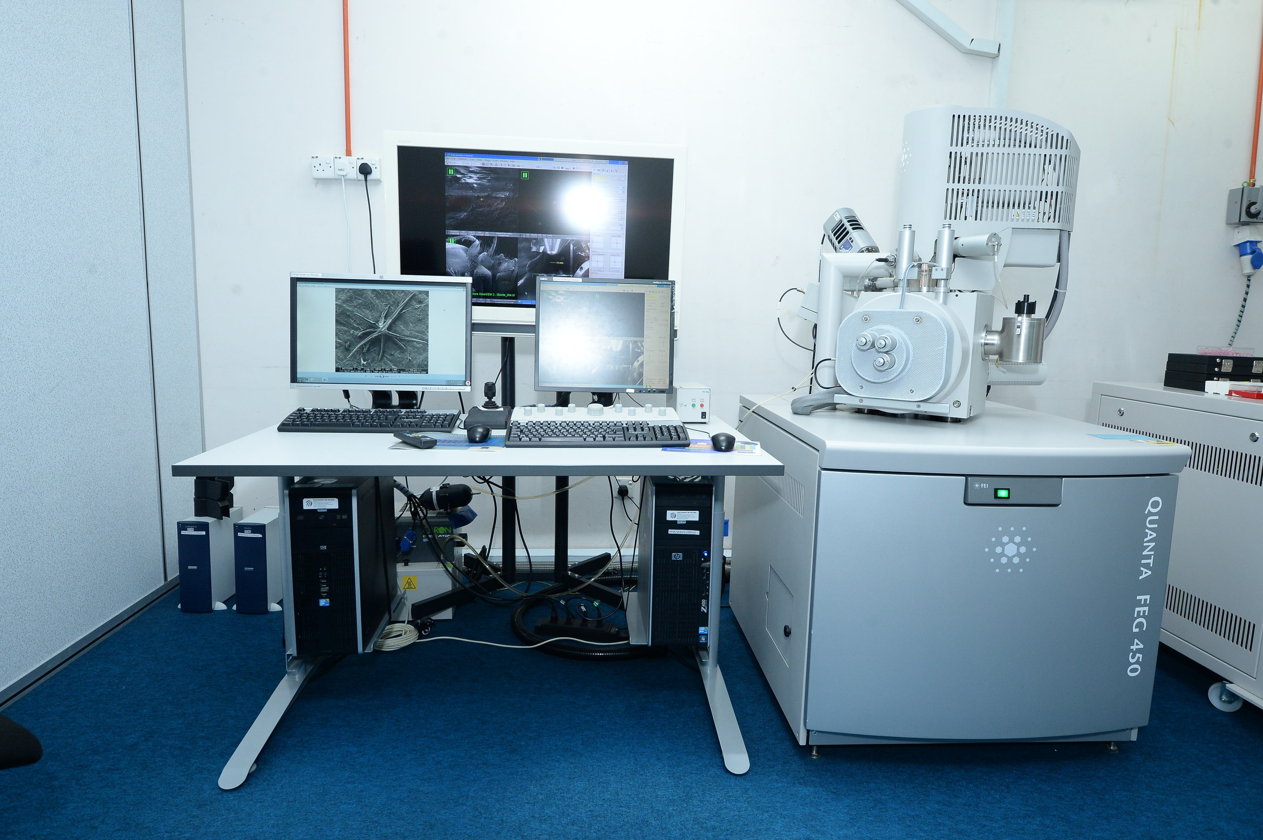
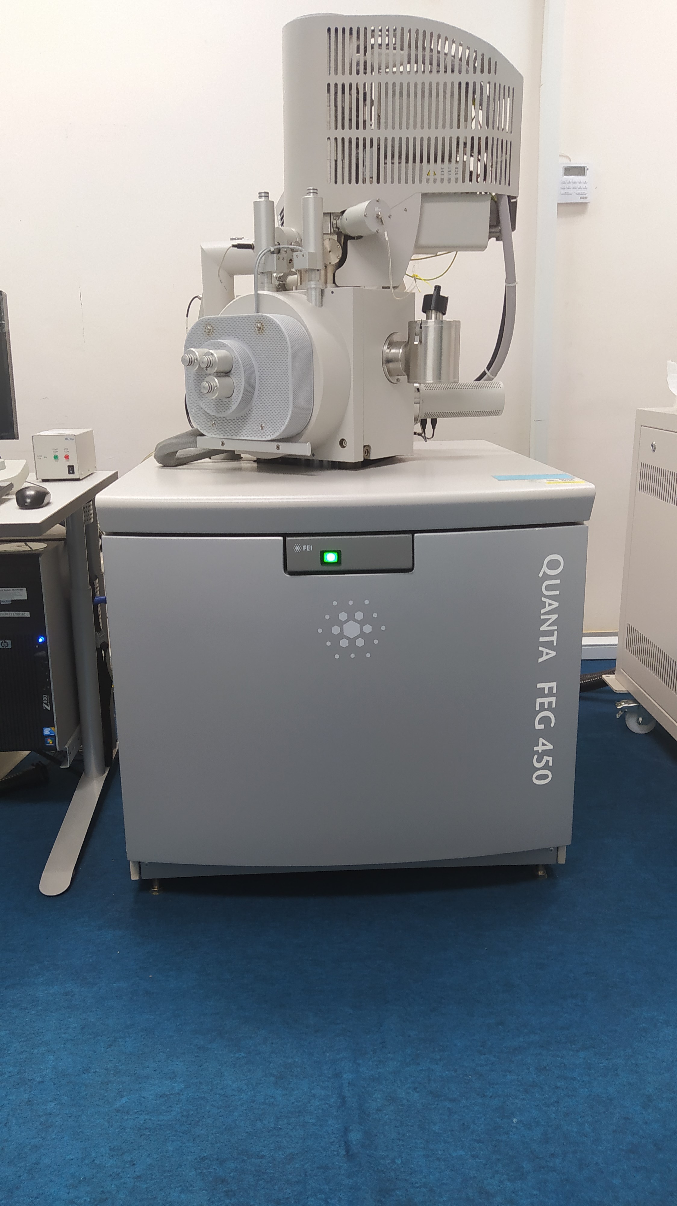
The FEI Quanta 450 FEG is a field emission gun – scanning electron microscope (FEG-SEM) used for high-resolution imaging (morphological and compositional) of both conductive and non-conductive specimens at the nanometer-scale resolution (magnification range: from 6 x to 1,000,000 x) and for semi-quantitative X-ray microanalysis. The instrument operates under three imaging modes: high and low vacuum and extended low vacuum modes. The instrument can thus accommodate multiple sample, imaging and analytical requirements for both material and life sciences, allowing investigation of a wide range of traditional and non-traditional samples with or without preparation, including wet samples in their natural state
- Manufacturer
- Czech Republic
- Brand
- FEI
- Model
- QUANTA FEG 450
- Year Manufactured
- Year Procured
- 2011
- Department
- PUSAT PENGAJIAN SAINS KESIHATAN
- Location
- Ppsk-blok E > Ground > Sem
- Date Registered LESIS
- 19/02/2024
- Category
- Research Equipment
- Function
- Booking, Testing,
- Category
- Staff operated
- Equipment Status
- Good
Person In-Charge

